
Whitening sensitive teeth can be safe with the right care. Learn how Rockwest Dental Clinic Mississauga customizes whitening treatments to protect enamel, reduce discomfort, and deliver bright results without added sensitivity.
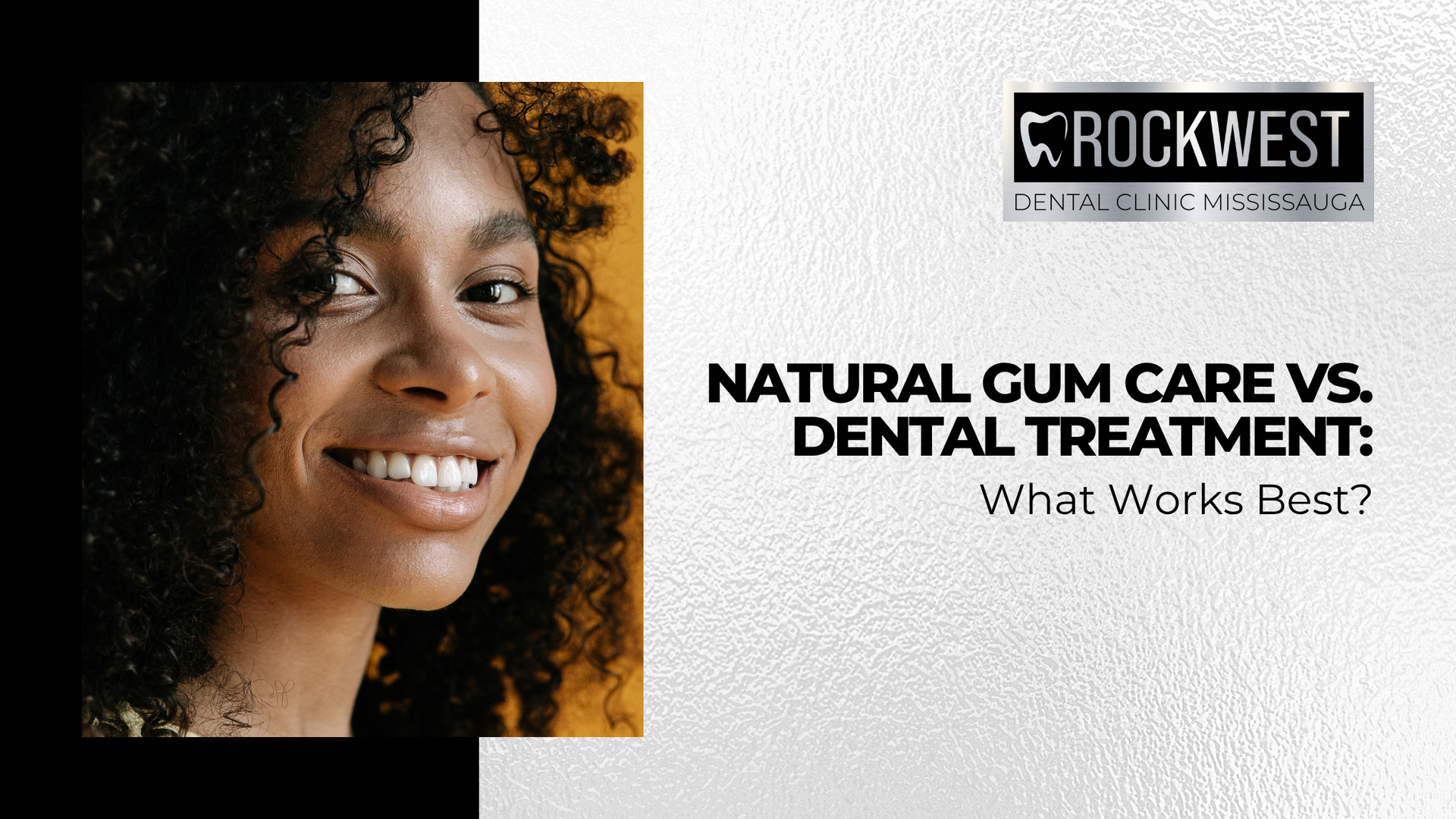
Natural remedies like saltwater rinses, oil pulling, and herbal treatments may offer short-term relief for gum irritation, but they are not a substitute for professional care. At Rockwest Dental Clinic Mississauga, we combine expert periodontal treatments with guidance on at-home care to help manage gum disease at every stage. Learn how to balance natural approaches with dental support to maintain healthy gums and prevent long-term oral health complications.
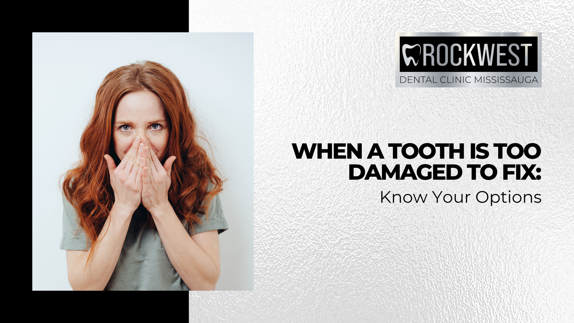
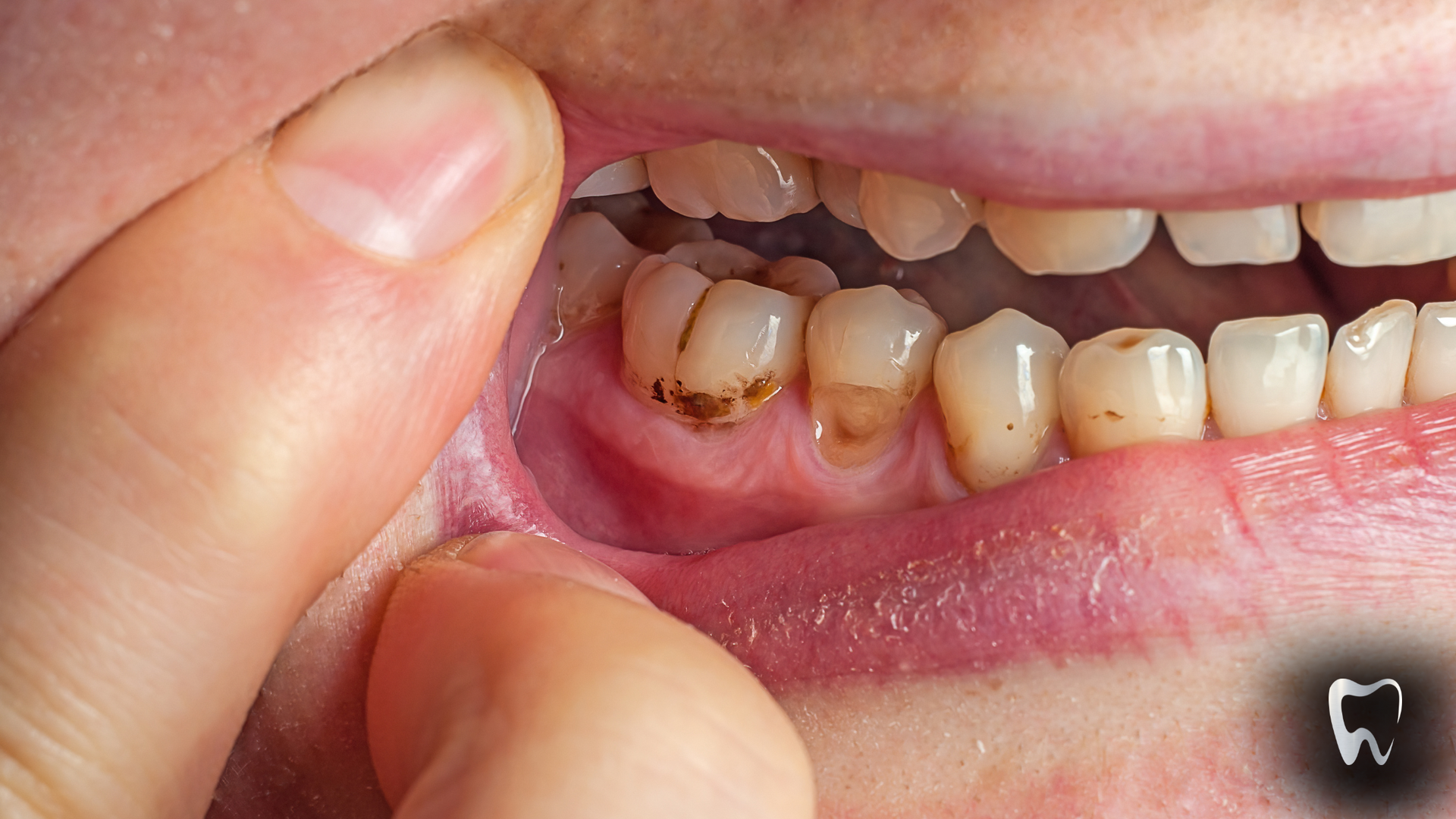
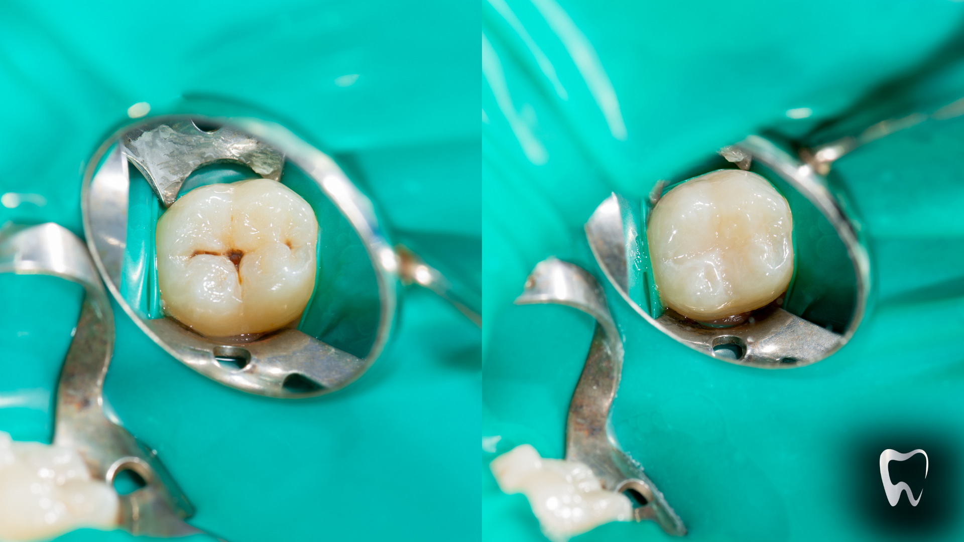
Is your tooth beyond saving? Rockwest Dental Clinic Mississauga outlines signs of severe damage, treatment paths for extraction, and modern replacement options like dental implants, bridges, and dentures. Learn how to identify serious symptoms early and how our team supports your oral health—from diagnosis to restoration. Start protecting your smile today with expert guidance and lasting solutions tailored to your needs.
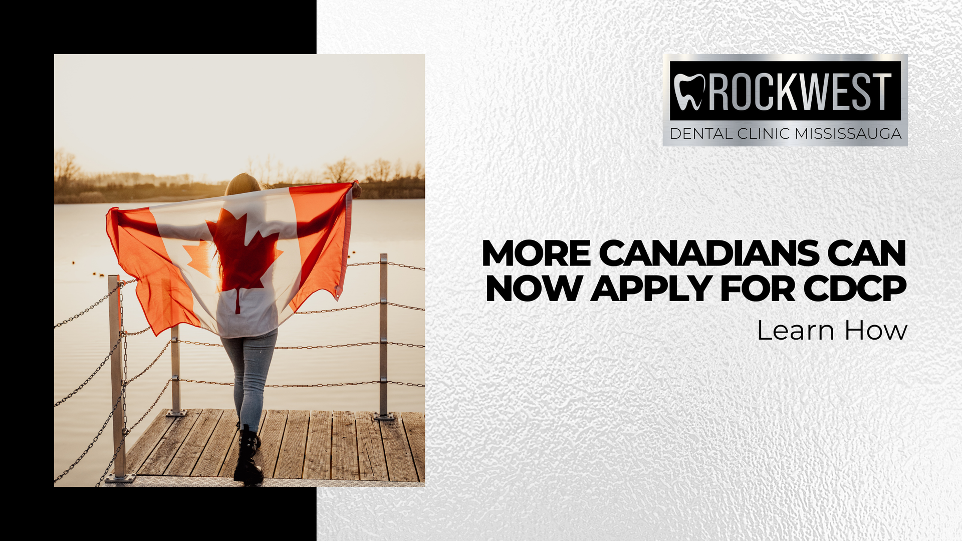
The Canadian Dental Care Plan (CDCP) has expanded application access as of May 2025, allowing more Canadians without dental insurance to apply for essential coverage. At Rockwest Dental Clinic Mississauga, we help patients navigate eligibility, co-pays, and available services. Learn how the CDCP works, who qualifies, and what to expect after applying—because understanding your benefits is the first step toward achieving better oral health and peace of mind.
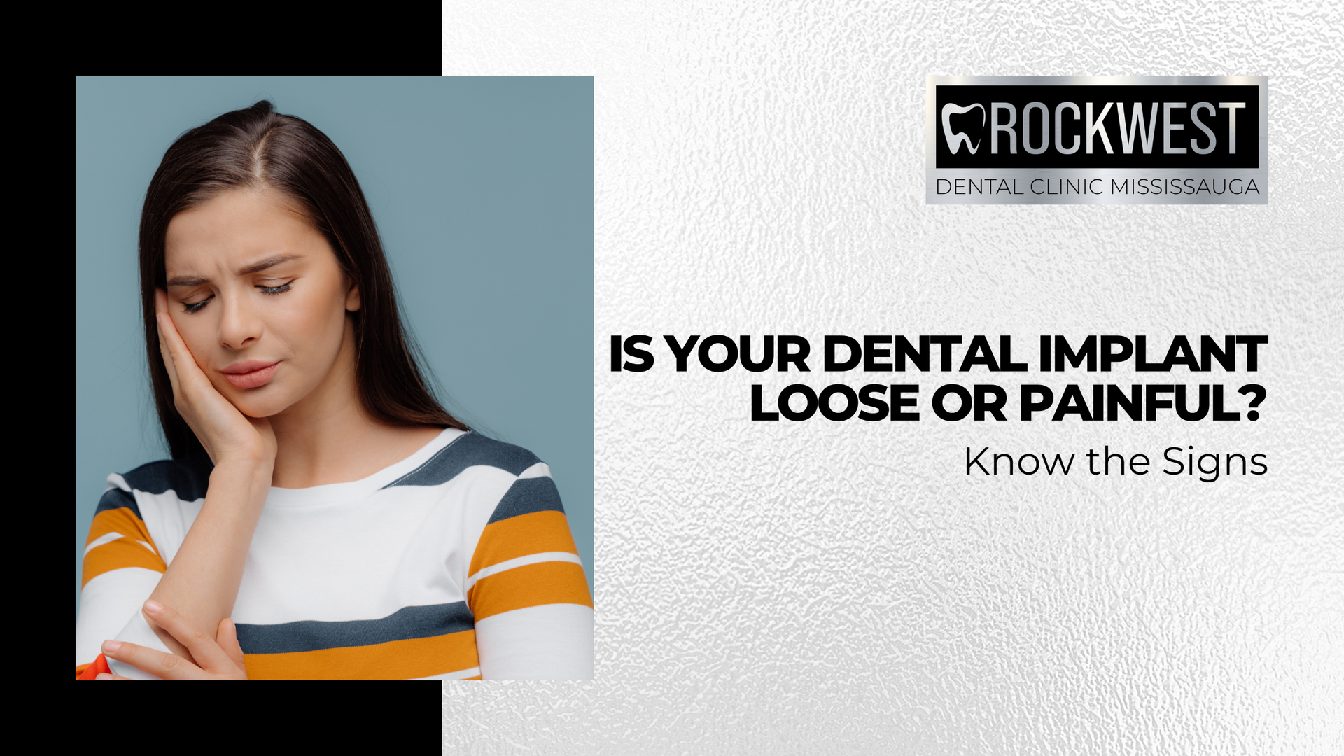
Dental implants are a durable solution, but issues like sensitivity, gum bleeding, or shifting can signal a problem. At Rockwest Dental Clinic Mississauga, we help patients identify red flags early and prevent long-term damage. Our team offers supportive care to ensure your implant remains secure and your smile stays strong.
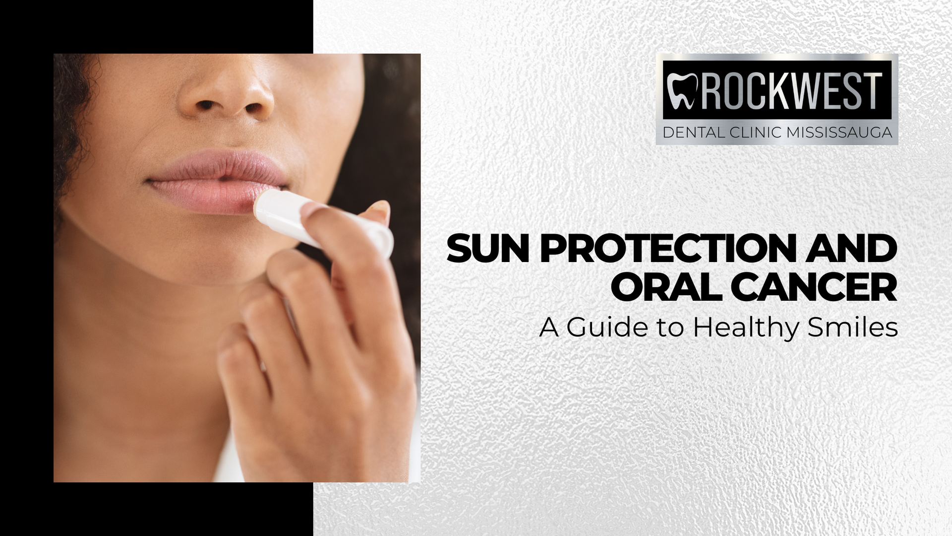
Prolonged sun exposure can damage delicate lip tissue, increasing oral cancer risk. At Rockwest Dental Clinic Mississauga, we emphasize prevention through SPF protection and regular screenings. Early detection is key to better health outcomes. Protect yourself with sun safety and proactive care. Book your oral cancer screening with us today and take a step toward a healthier future!
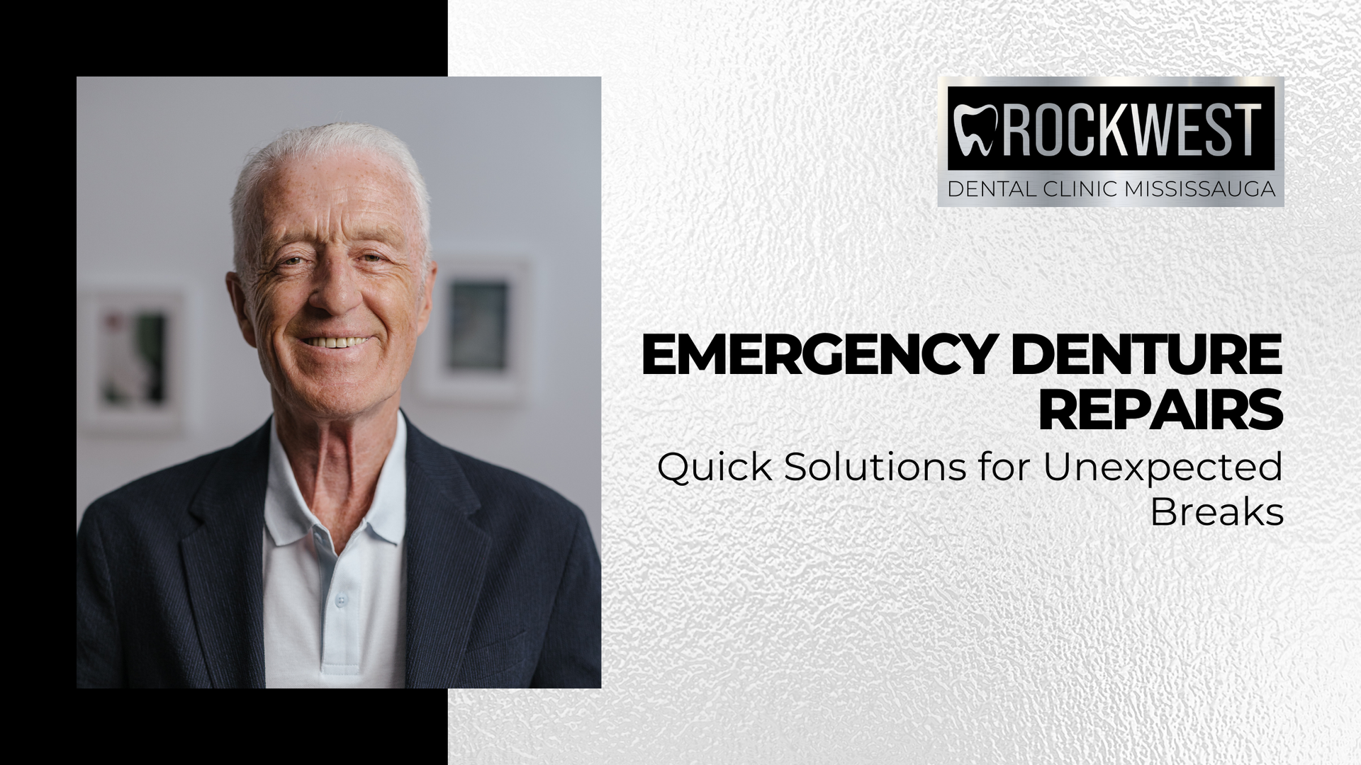
Broken dentures? Do not panic! At Rockwest Dental Clinic Mississauga, we specialize in emergency denture repairs to get your smile back in no time. Whether your dentures cracked, chipped, or feel loose, our team offers quick, effective solutions to restore comfort and function. Avoid DIY fixes—book your denture repair with us today and get back to smiling confidently!
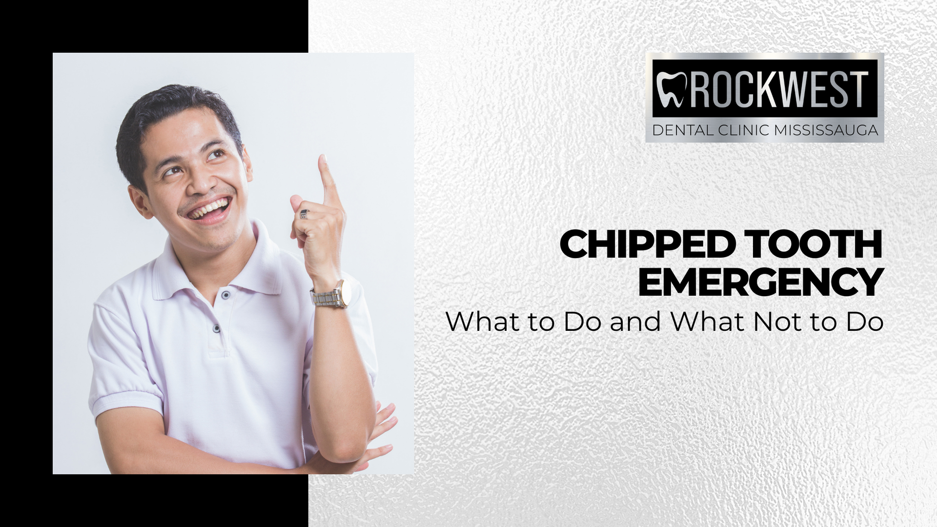
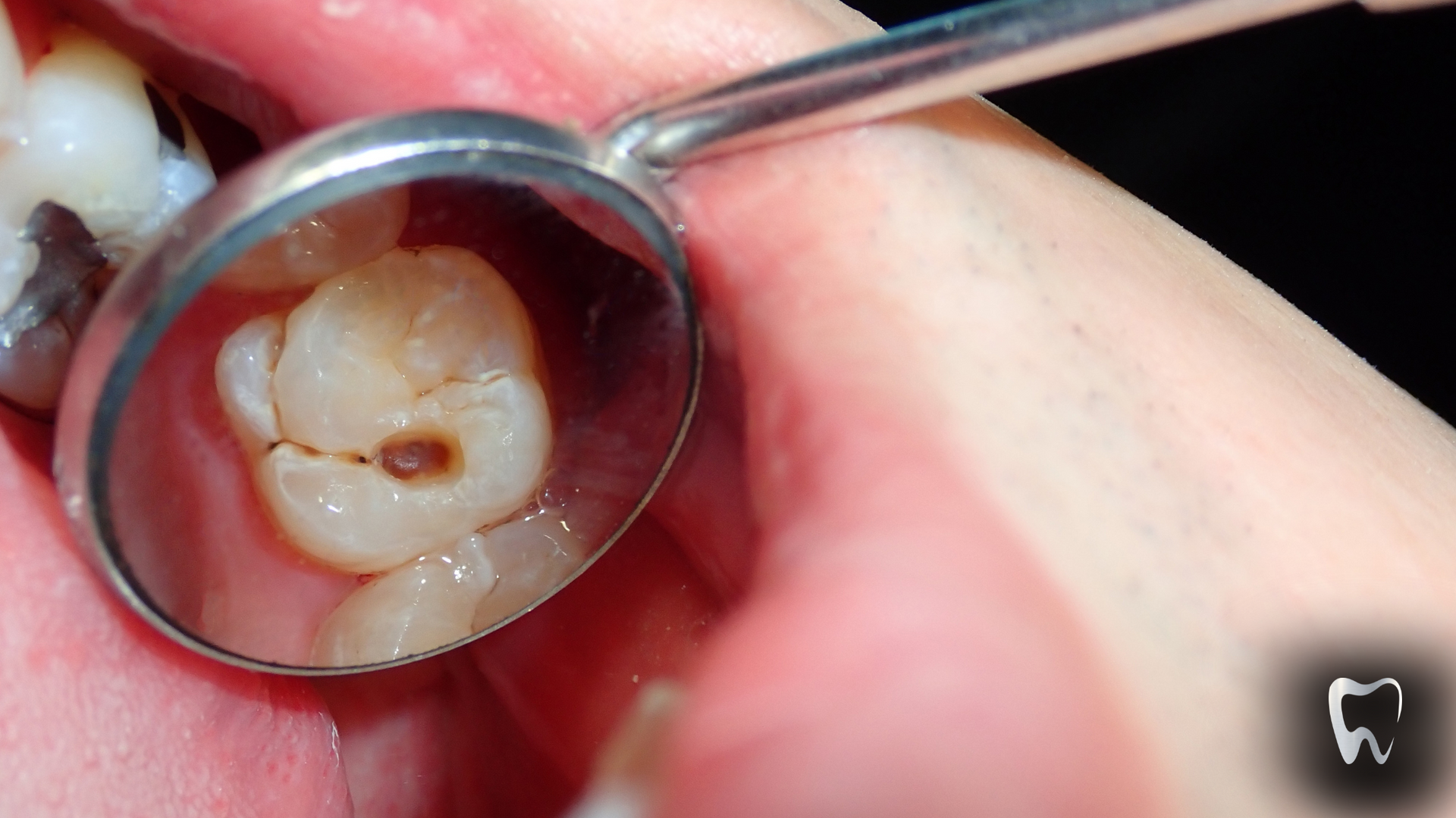
Wondering if you can fix a chipped tooth at home? Discover why DIY repairs might do more harm than good and learn why professional dental care is crucial for a chipped tooth. At Rockwest Dental Clinic Mississauga, we provide safe, effective emergency treatments to restore your smile. Avoid the risks of home remedies—contact us today for expert care!
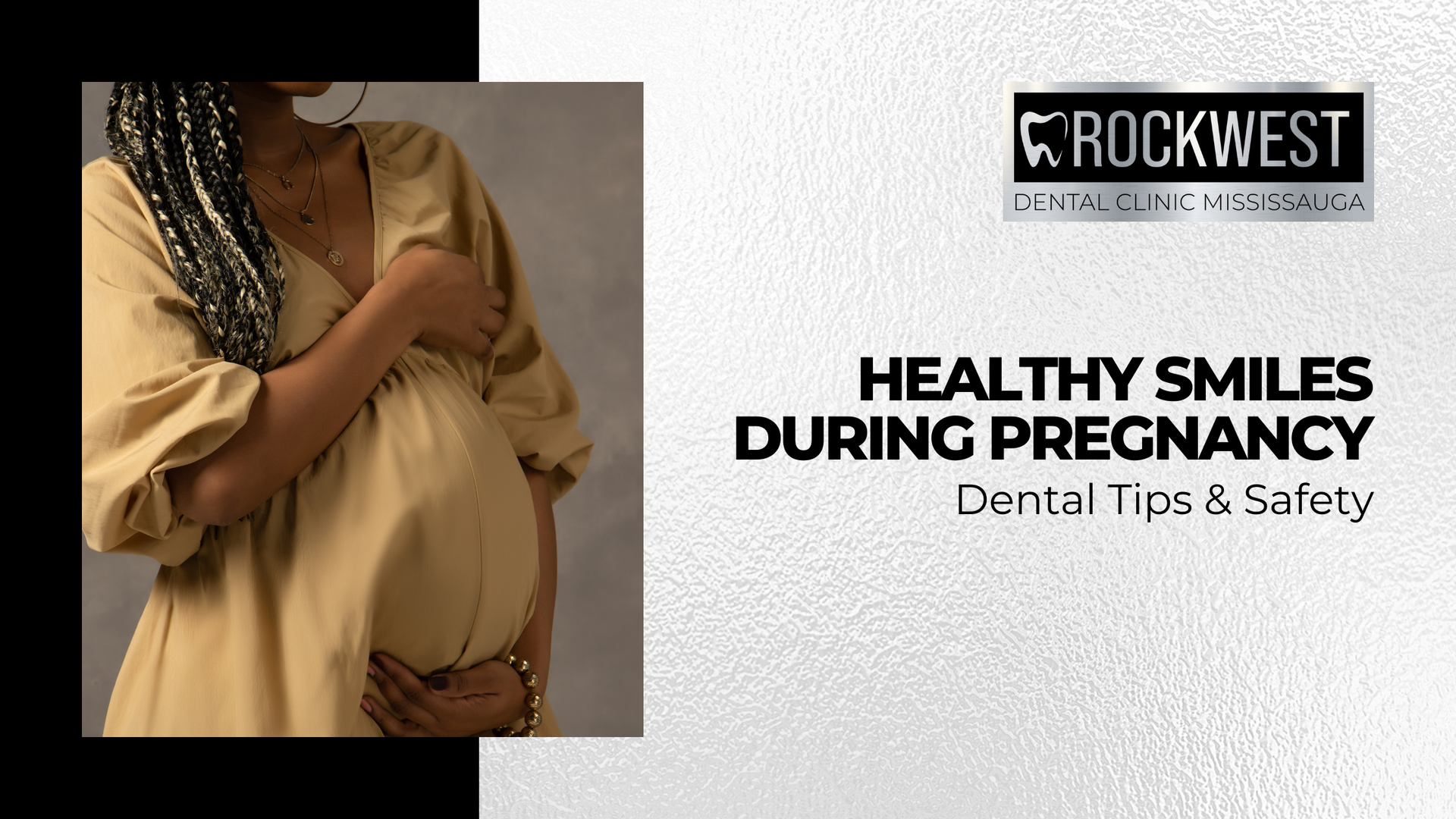
Good oral health is essential during pregnancy. Hormonal changes can lead to gum inflammation, cavities, and enamel erosion. Discover safe dental treatments and habits to maintain a healthy smile while protecting your baby. At Rockwest Dental Clinic Mississauga, we offer safe, compassionate dental care for expectant individuals.

Dental abscesses can lead to severe health risks if ignored. At Rockwest Dental Clinic Mississauga, our expert team offers personalized treatment to eliminate infections and prevent complications like sinusitis and sepsis. Take control of your oral health—book your visit today!

Whitening sensitive teeth can be safe with the right care. Learn how Rockwest Dental Clinic Mississauga customizes whitening treatments to protect enamel, reduce discomfort, and deliver bright results without added sensitivity.
Natural remedies like saltwater rinses, oil pulling, and herbal treatments may offer short-term relief for gum irritation, but they are not a substitute for professional care. At Rockwest Dental Clinic Mississauga, we combine expert periodontal treatments with guidance on at-home care to help manage gum disease at every stage. Learn how to balance natural approaches with dental support to maintain healthy gums and prevent long-term oral health complications.
Is your tooth beyond saving? Rockwest Dental Clinic Mississauga outlines signs of severe damage, treatment paths for extraction, and modern replacement options like dental implants, bridges, and dentures. Learn how to identify serious symptoms early and how our team supports your oral health—from diagnosis to restoration. Start protecting your smile today with expert guidance and lasting solutions tailored to your needs.